Dural arteriovenous fistula
| Dural arteriovenous fistula | |
|---|---|
 | |
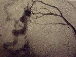
| This dural arteriovenous fistula of the superior sagittal sinus drains into subarachnoid veins and is classified as Borden type IIIb. | |
| Classification and external resources | |
| DiseasesDB | 32954 |
A dural arteriovenous fistula (DAVF), is an abnormal direct connection (fistula) between a meningeal artery and a meningeal vein or dural venous sinus. In cases where there are multiple fistulas, the related term dural arteriovenous malformation (DAVM) is used.
Classification
Borden Classification
The Borden Classification of dural arteriovenous malformations or fistulas, groups into three types based upon their venous drainage:[1]
- Type I: dural arterial supply drains anterograde into venous sinus.
- Type II: dural arterial supply drains into venous sinus. High pressure in sinus results in both anterograde drainage and retrograde drainage via subarachnoid veins.
- Type III: dural arterial supply drains retrograde into subarachnoid veins.
Type I
Type I dural arteriovenous fistulas are supplied by meningeal arteries and drain into a meningeal vein or dural venous sinus. The flow within the draining vein or venous sinus is anterograde.
- Type Ia – simple dural arteriovenous fistulas have a single meningeal arterial supply
- Type Ib – more complex arteriovenous fistulas are supplied by multiple meningeal arteries
The distinction between Types Ia and Ib is somewhat specious as there is a rich system of meningeal arterial collaterals. Type I dural fistulas are often asymptomatic, do not have a high risk of bleeding and do not necessarily need to be treated.
Type II
The high pressure within a Type II dural AV fistula causes blood to flow in a retrograde fashion into subarachnoid veins which normally drain into the sinus. Typically this is because the sinus has outflow obstruction. Such draining veins form venous varices or aneurysms which can bleed. Type II fistulas need to be treated to prevent hemorrhage. The treatment may involve embolization of the draining sinus as well as clipping or embolization of the draining veins.
Type III
Type III dural AV fistulas drain directly into subarachnoid veins.[2] These veins can form aneurysms and bleed. Type III dural fistulas need to be treated to prevent hemorrhage. Treatment can be as simple as clipping the draining vein at the site of the dural sinus. If treatment involves embolization, it will only typically be effective if the glue traverses the actual fistula and enters, at least slightly, the draining vein.
Cognard Classification
The Cognard Classification[3] correlates venous drainage patterns with increasingly aggressive neurological clinical course.
Type I
Confined to sinus wall, typically after thrombosis.
Type II
IIa - confined to sinus with reflux (retrograde) into sinus but not cortical veins. IIb - drains into sinus with reflux (retrograde) into cortical veins (10-20% hemorrhage).
Type III
Drains direct into cortical veins (not into sinus) drainage (40% hemorrhage).
Type IV
Drains direct into cortical veins (not into sinus) drainage with venous ectasia (65% hemorrhage).
Type V
Spinal perimedullary venous drainage, associated with progressive myelopathy.
Treatment
One approach used for treatment is embolization.[4]
See also
References
- ↑ Borden JA, Wu JK, Shucart WA (1995). "A proposed classification for spinal and cranial dural arteriovenous fistulous malformations and implications for treatment". J. Neurosurg. 82 (2): 166–79. doi:10.3171/jns.1995.82.2.0166. PMID 7815143.
- ↑ "jonathanborden-md.com". Retrieved 2007-12-22.
- ↑ Cognard C, Gobin YP, Pierot L et-al. Cerebral dural arteriovenous fistulas: clinical and angiographic correlation with a revised classification of venous drainage. Radiology. 1995;194 (3): 671-80.
- ↑ Carlson AP, Taylor CL, Yonas H (2007). "Treatment of dural arteriovenous fistula using ethylene vinyl alcohol (onyx) arterial embolization as the primary modality: short-term results". J. Neurosurg. 107 (6): 1120–5. doi:10.3171/JNS-07/12/1120. PMID 18077948.