Bilateral hilar lymphadenopathy
Bilateral hilar lymphadenopathy is a bilateral enlargement of the lymph nodes of pulmonary hila. It is a radiographic term that describes the enlargement of mediastinal lymph nodes and is most commonly identified by a chest x-ray.

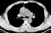
CT scan of the chest showing bilateral lymphadenopathy in the mediastinum due to sarcoidosis.
Causes
The following are causes of BHL:[1]
- Sarcoidosis[2]
- Infection
- Tuberculosis[2]
- Fungal infection[2]
- Mycoplasma
- Intestinal Lipodystrophy (Whipple's disease)[3][4]
- Malignancy
- Inorganic dust disease
- Extrinsic allergic alveolitis
- Such as bird fancier's lung
- Less common causes also exist:
References
- ↑ M. Longmore; I. Wilkinson; T. Turmezei; CK. Cheug (2007). Oxford Handbook of Clinical Medicine 7th Edition. United States, New York: Oxford University Press. p. 179. ISBN 0-19-856837-1.
- 1 2 3 4 Criado, E; Sánchez, M; Ramírez, J; Arguis, P; De Caralt, TM; Perea, RJ; Xaubet, A (October 2010). "Pulmonary sarcoidosis: typical and atypical manifestations at high-resolution CT with pathologic correlation". Radiographics. 30 (6): 1567–1586. doi:10.1148/rg.306105512. PMID 21071376.
- ↑ Urbanksi, G; Rivereau, P; Artru, L; Fenollar, F; Raoult, D; Puéchal, X (June 2012). "Whipple disease revealed by lung involvement: a case report and literature review". Chest. 141 (6): 1595–1598. doi:10.1378/chest.11-1812. PMID 22670021.
- ↑ Beers, Mark (2006). The Merck Manual.
- ↑ Suwatanapongched, T; Gierada, DS (December 2006). "CT of thoracic lymph nodes. Part II: diseases and pitfalls". The British Journal of Radiology. 79 (948): 999–1000. doi:10.1259/bjr/82484604. PMID 16641412.
- ↑ Leung, CC; Yu, IT; Chen, W (May 2012). "Silicosis". The British Journal of Radiology. 379 (9830): 2008–2018. doi:10.1016/S0140-6736(12)60235-9. PMID 22534002.
- 1 2 Chong, S; Lee, KS; Chung, MJ; Han, J; Kwon, OJ; Kim, TS (January–February 2006). "Pneumoconiosis: comparison of imaging and pathologic findings". Radiographics. 26 (1): 59–77. doi:10.1148/rg.261055070. PMID 16418244.
- ↑ Owlia, MB; Mehrpoor, G (May 2009). "Adult-onset Still's disease: a review". Indian Journal of Medical Sciences. 63 (5): 207–221. doi:10.4103/0019-5359.53169. PMID 19584494.
This article is issued from Wikipedia - version of the 7/16/2016. The text is available under the Creative Commons Attribution/Share Alike but additional terms may apply for the media files.